For people with a type of jaw joint disorder that results from loss of cartilage, the only treatments available address symptoms but do not repair the damaged tissue. Now, a new study of mice suggests that stem cells already present in the jaw joint could be manipulated to repair it.
Columbia University Medical Center in New York led the study and the journal Nature Communications published it. The authors describe how manipulating stem cells in the temporomandibular joint (TMJ) of mice with TMJ degeneration led the cells to repair cartilage in the joint. They also found that transplanting a single TMJ stem cell into a mouse spontaneously generated cartilage and bone and began to form a bone marrow niche.
Lead corresponding author Mildred C. Embree, DMD, Ph.D., assistant professor of dental medicine at Columbia, said, “This is very exciting for the field because patients who have problems with their jaws and TMJs are very limited in terms of clinical treatments available.”
“Stem cells—immature cells that have the potential to mature into virtually any type of tissue cell—hold great promise for regenerative medicine, where faulty, damaged, or injured tissue is repaired by encouraging new cells to grow.”
One method of regenerating tissue is to transplant stem cells into the affected area. As the authors explain, one way to generate tissue is to transplant stem cells into the affected areas. However, as the authors explain in their paper, this approach can be risky as donor cells can be rejected by the recipient’s immune system, can introduce pathogens and give rise to tumors.
An alternative is that stem cells already present in the affected area could be induced to make new cells to repair the damaged tissue.
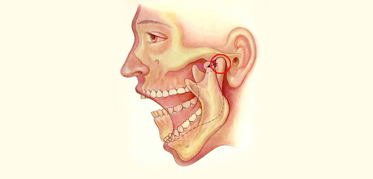
Within the TMJ, the cartilage is of a type called fibrocartilage. This type is also found in the knee meniscus and the discs between the vertebrae of the spine.
Once it is damaged through injury or disease—fibrocartilage does not regrow or heal, with the result that people suffer permanent disability.
In the United States alone, there are up to 10 million people—mostly women—with TMJ disorders. Children with juvenile idiopathic arthritis can also have stunted jaw growth that cannot be treated with existing drugs. The researchers suggest their findings could lead to new treatments that help these groups.
In their study, Professor Embree and colleagues—including Jeremy Mao, DDS, Ph.D., the Edwin S. Robinson Professor of Dentistry (in Orthopedic Surgery) at Columbia—show for the first time that the “fibrous superficial zone” in the TMJ of mice harbors fibrocartilage stem cells (FCSCs).
They also discovered that a single FCSC transplanted into a live mouse is capable of not only generating cartilage and bone, but also of organizing the microenvironment to support the process.
Moreover, the researchers showed that they could manipulate FCSCs to differentiate into the required cell types by suppressing a type of cell signal called Wnt. They found that overactive Wnt signals disrupt the stability of fibrocartilage and promote degeneration by depleting FCSCs.
The findings could also open up routes to new treatments for repairing fibrocartilage in joints other than the TMJ, including the knees and vertebral discs, as Professor Embree explains: “Those types of cartilage have different cellular constituents, so we would have to really investigate the molecular underpinnings regarding how these cells are regulated. The implications of these findings are broad. They suggest that molecular signals that govern stem cells may have therapeutic applications for cartilage and bone regeneration.”
